Things to know about uterine fibroids
What are uterine fibroids?
Uterine fibroids are benign tumors that originate in the uterus (womb). It is also called an Uterina myoma.
- It is not known exactly why women develop uterine fibroids.
- Most women with uterine fibroids have no symptoms. However, fibroids can cause a number of symptoms depending on their size, location within the uterus, and how close they are to adjacent pelvic organs. These are most commonly abnormal bleeding, pain, and pressure.
- Uterine fibroids are diagnosed by pelvic exam and by ultrasound.
- If treatment for uterine fibroids is necessary, several options are available that include surgery (hysterectomy, myomectomy, cryosurgery), MRI-guided high-intensity focused ultrasound (MRgFUS or HIFU), and uterine artery embolization (UAE).
- Medical treatments include medications such as mifepristone (RU-486), danazol (Danocrine), raloxifene (Evista), GnRH analogs (Lupron and others), and low-dose formulations of oral contraceptives.
- Estrogen tends to stimulate the growth of fibroids in many cases.

Uterine Fibroids Trreatment
Why is a hysterectomy performed?
The most common reason hysterectomy is performed is for uterine fibroids. Other common reasons are:
- abnormal uterine bleeding (vaginal bleeding),
- cervical dysplasia (pre-cancerous conditions of the cervix),
- endometriosis, and uterine prolapse (including pelvic relaxation).

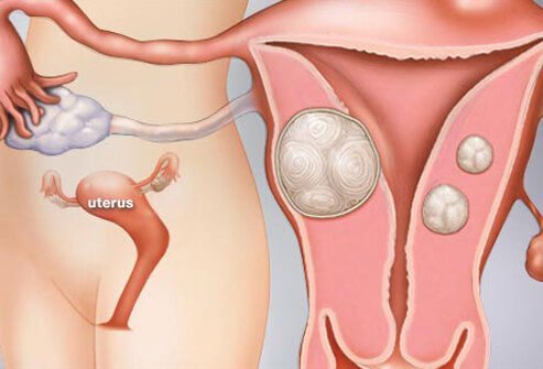
What are uterine fibroids? What do they look like?
Picture of uterine fibroids
Uterine fibroids are benign tumors that originate in the uterus (womb). Although they are composed of the same smooth muscle fibers as the uterine wall (myometrium), they are much denser than normal myometrium. Uterine fibroids are usually round. In most cases, fibroids do not cause pain or other symptoms. However, exceptionally large fibroids may cause pressure on the bladder or other organs, leading to specific symptoms (see: What are the symptoms of uterine fibroids?)
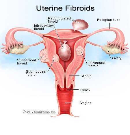
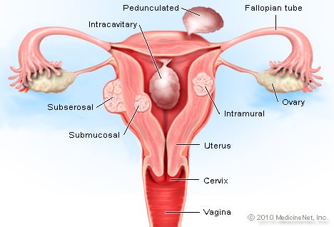
Uterine fibroids are often described based on their location within the uterus.
- Subserosal fibroids are located beneath the serosa (the lining membrane on the outside of the uterus). These often appear localized on the outside surface of the uterus or may be attached to the outside surface by a pedicle.
- Submucosal (submucous) fibroids are located inside the uterine cavity beneath the inner lining of the uterus.
- Intramural fibroids are located within the muscular wall of the uterus.
- Pedunculated fibroids grow on a stalk of tissue known as a pedicle (like a mushroom), extending either inside the cavity of the uterus or outside the uterus from its outer surface.
SLIDESHOW
What Are Uterine Fibroids? Symptoms, Treatment, Pictures See Slideshow
What are the symptoms of uterine fibroids? Do they cause pain?
Most of the time, uterine fibroids do not cause symptoms or problems, and a woman with a fibroid is usually unaware of its presence.
However, abnormal uterine bleeding is the most common symptom of a fibroid. If the tumors are near the uterine lining or interfere with the blood flow to the lining, they can cause heavy periods, painful periods, prolonged periods, or spotting between menses. Women with excessive bleeding due to fibroids may develop iron deficiency anemia. Uterine fibroids that are degenerating can sometimes cause severe, localized pain.
Fibroids can also cause a number of symptoms depending on their size, location within the uterus, and how close they are to adjacent pelvic organs. Large fibroids can cause:
- pressure,
- pelvic pain, including pain during sex,
- pressure on the bladder with frequent or even obstructed urination, and
- pressure on the rectum with painful or difficult defecation.
Uterine fibroids and pregnancy
While fibroids do not interfere with ovulation, some studies suggest that they may impair fertility and lead to poorer pregnancy outcomes. In particular, submucosal fibroids that deform the inner uterine cavity are most strongly associated with decreases in fertility. Occasionally, fibroids are the cause of recurrent miscarriages. If they are not removed in these cases, the woman may not be able to sustain a pregnancy.
Latest Women’s Health News
Daily Health News
Trending on MedicineNet
What causes uterine fibroids to grow? How big can they get?
We do not know exactly why women develop these tumors. Genetic abnormalities, alterations in growth factor (proteins formed in the body that direct the rate and extent of cell proliferation) expression, abnormalities in the vascular (blood vessel) system, and tissue response to injury have all been suggested to play a role in the development of fibroids.
Family history is a key factor since there is often a history of fibroids developing in women of the same family. The race also appears to play a role. Women of African descent are two to three times more likely to develop fibroids than women of other races. Women of African ancestry also develop fibroids at a younger age and may have symptoms from fibroids in their 20s, in contrast to Caucasian women with fibroids, in whom symptoms typically occur during the 30s and 40s. Early pregnancy decreases the likelihood that fibroids will develop. Fibroids have not been observed in girls who have not reached puberty, but adolescent girls may rarely develop fibroids.
Other factors that researchers have associated with an increased risk of developing fibroids include
Estrogen tends to stimulate the growth of fibroids in many cases. During the first trimester of pregnancy, about a third of fibroids will enlarge and then shrink after the birth. In general, fibroids tend to shrink after menopause, but postmenopausal hormone therapy may cause symptoms to persist.
Overall, these tumors are fairly common and occur in about 70% to 80% of all women by the time they reach age 50.
Uterine fibroids can be as small as a few millimeters (less than an inch) in diameter. They can also be very large (grapefruit-sized or larger).
IMAGES
Uterine Fibroids (Benign Tumors of the Uterus) See a medical illustration of uterine fibroids plus our entire medical gallery of human anatomy and physiology See Images
Are fibroids serious? Can fibroids disappear on their own?
For the most part, uterine fibroids that do not cause a problem for the woman can be left untreated. In some cases, even fibroids that are not causing symptoms require removal or at least close observation. Rapid growth is a reason to watch more carefully, since a rare cancerous form of fibroid (referred to as a leiomyosarcoma) can be a fast-growing tumor, and it cannot be differentiated from a benign fibroid by ultrasound, MRI, or other imaging studies. However, this type of tumor occurs in less than 1% of uterine fibroids. It is also important to note that these rare cancerous tumors are not thought to begin in a benign fibroid.
Another risk of leaving these tumors alone is that they sometimes grow to a size that eventually causes significant symptoms, thus requiring removal. If fibroids grow large enough, the surgery to remove them can become more difficult and risky.
Subscribe to MedicineNet’s Women’s Health Newsletter
By clicking “Submit,” I agree to the MedicineNet Terms and Conditions and Privacy Policy. I also agree to receive emails from MedicineNet and I understand that I may opt out of MedicineNet subscriptions at any time.
What tests diagnose uterine fibroids?
Uterine fibroids are diagnosed by pelvic exam and even more commonly by ultrasound. Often, a pelvic mass cannot be determined to be a fibroid on a pelvic exam alone, and ultrasound is very helpful in differentiating it from other conditions such as ovarian tumors.
- MRI and CT scans can also play a role in diagnosing fibroids, but ultrasound is the simplest, cheapest, and best technique for imaging the pelvis.
- Occasionally, when trying to determine if a fibroid is present in the uterine cavity (endometrial cavity), a hysterosonogram (HSG) is done. In this procedure, an ultrasound exam is done while contrast fluid is injected into the uterus through the cervix. The fluid within the endometrial cavity can help outline any masses that are inside, such as submucosal fibroids.
Are there home remedies for uterine fibroids?
There are no known home remedies that can shrink fibroids. If uterine fibroids are not causing symptoms or problems, they can be left alone without specific treatment.
- If they are large enough to cause symptoms like bleeding, pain, or pressure, medical or surgical treatment is required.
What is the treatment for uterine fibroids?
There are several uterine fibroids treatment options, including:
- surgery (hysterectomy, myomectomy, cryosurgery),
- MRI-guided high-intensity focused ultrasound (MRgFUS), and
- uterine artery embolization (UAE).
Medical treatments include medications such as:
- mifepristone (RU-486),
- danazol (Danocrine),
- raloxifene (Evista),
- GnRH analogs (Lupron and others), and
- low-dose formulations of oral contraceptives.
From 
Women’s Health Resources
Featured Centers
Health Solutions From Our Sponsors
Surgery for fibroids
There are many ways of managing uterine fibroids. Surgical methods are the mainstay of treatment when treatment is necessary. Possible surgical interventions include
- hysterectomy or removal of the uterus (and the fibroids with it).
- Myomectomy is the selective removal of just the fibroids within the uterus.
- Myomectomy can be done through a hysteroscope, laparoscope, or with the standard open incision on the abdominal wall.
- Some treatments have involved boring holes into the fibroid with laser fibers, freezing probes (cryosurgery), and other destructive techniques that do not actually remove the tissue but try to destroy it in place.
- Surgery is necessary if there is suspicion of malignancy in any case of a leiomyoma or uterine mass.
Another technique for treating fibroids is known as uterine artery embolization (UAE). This technique uses small beads of a compound called polyvinyl alcohol, which is injected through a catheter into the arteries that feed the fibroid. These beads obstruct the blood supply to the fibroid and starve it of blood and oxygen. While this technique has not been in use long enough to evaluate the long-term effects of UAE versus surgery, women undergoing UAE for fibroids have a shorter hospital stay than those having surgery but a greater risk of complications and readmissions to the hospital. Studies are underway to evaluate the long-term outcomes of UAE as opposed to surgical treatment.
Uterine artery occlusion (UAO), which involves clamping the involved uterine arteries as opposed to injecting the polyvinyl alcohol beads, is currently under investigation as a potential alternative to UAE.
High-intensity focused ultrasound (HIFU) is a relatively new treatment for fibroids and other abnormalities. It is also known as MRgFUS (MRI-guided focused ultrasound) and FUS (focused ultrasound surgery). HIFU uses an ultrasound transducer with higher energy than those used for diagnostic examinations. The device focuses on the sound waves, generating heat to destroy the fibroid. MRI imaging may be used for planning and monitoring treatment.
Medical treatment for fibroids
Nonsurgical techniques are usually hormonal in nature and include the use of drugs that turn off the production of estrogen from the ovaries (GnRH analogs). These medications are given for three to six months and induce a hypoestrogenic (low estrogen) state. When successful, they can shrink the fibroids by as much as 50%. Side effects of these drugs are similar to the symptoms of menopause and can include hot flashes, sleep disturbance, vaginal dryness, and mood changes. Bone loss leading to osteoporosis after long-term (6 to 12+ months) use is one complication. This is generally reversed after the treatment ends. These drugs may also be used as preoperative treatment for large leiomyomas to shrink them in order to make the operation less difficult and reduce surgical risk.
Mifepristone (RU-486) is an antiprogestin drug that can shrink fibroids to an extent comparable to treatment with the GnRH analogs. This drug is also used to terminate early pregnancy. Treatment with mifepristone also reduces the bleeding associated with fibroids, but this treatment can be associated with adverse side effects such as overgrowth (hyperplasia) of the endometrium (uterine lining). Mifepristone is not approved by the US Food and Drug Administration (FDA) for the treatment of uterine leiomyomas, and the required dosages (different from those used for termination of early pregnancy) have not been determined.
Danazol (Danocrine) is an androgenic steroid hormone that has been used to reduce bleeding in women with fibroids since this drug causes menstruation to cease. However, danazol does not appear to shrink the size of fibroids. Danazol is also associated with significant side effects, including weight gain, muscle cramps, decreased breast size, acne, hirsutism (inappropriate hair growth), oily skin, mood changes, depression, decreased high-density lipoprotein (HDL or “good” cholesterol) levels, and increased liver enzyme levels.
The administration of raloxifene (Evista), a drug used to prevent and treat osteoporosis in postmenopausal women, has been shown to decrease the size of fibroids in postmenopausal women, but the results of this therapy in premenopausal women have been conflicting.
Low-dose formulations of oral contraceptives are also sometimes given to treat the abnormal bleeding associated with fibroids, but these do not shrink the fibroids themselves.
What are the risks of uterine fibroids during pregnancy?
Some studies have shown an increased risk of pregnancy complications in the presence of fibroids, such as first trimester bleeding, breech presentation, placental abruption, and problems during labor.
Fibroids have also been associated with an increased risk of cesarean delivery. The size of the fibroid and its precise location within the uterus are important factors in determining whether a fibroid causes obstetric complications.
Medically Reviewed on 4/28/2022
References
El-Balat, A., et al. “Modern Myoma Treatment in the Last 20 Years: A Review of the Literature.” BioMed Research International. 2018. <https://www.hindawi.com/journals/bmri/2018/4593875/>.
Office of Women’s Health, U.S. Department of Health and Human Services. “Uterine Fibroids.” April 1, 2019. <https://www.womenshealth.gov/a-z-topics/uterine-fibroids>.
Parker, W.H. “Uterine myomas: management.” Fertil Steril. 88.2 August 2007: 255-71. Epub July 20, 2007. <https://www.fertstert.org/article/S0015-0282(07)01408-2/fulltext>.
Thomason, P. “Uterine Leiomyoma (Fibroid) Imaging.” Medscape. Oct. 8, 2017. <http://emedicine.medscape.com/article/405676-overview>.