The healing process for radius fractures depends upon certain factors such as the following.
The radius is one of two forearm bones that is located on the thumb side (lateral aspect) of the forearm.
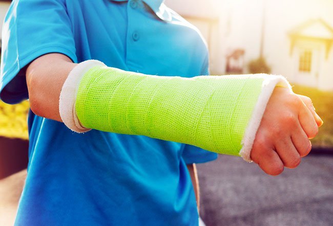
The radius fracture may be near the elbow (proximal) or the wrist (distal). Most distal radius fractures take about three months or more to heal before you can return to all activities. Some residual soreness and stiffness may take up to one year or even more.
Proximal radius fracture heals faster in around 6 to 12 weeks.
Factors that influence recovery time from a radial fracture
The healing from fractures depends upon the following factors:
- Age: Fractures heal faster and better in kids as compared to the elderly.
- The severity of trauma: A fracture in which the bone is crushed into multiple pieces takes more time to heal.
- Osteoporosis: Fractures in the elderly may not heal at all or take longer to heal due to poor bone health.
- Treatment approach: Open surgery has a poorer success rate compared to endoscopic approaches.
- Type of fracture: Fractures involving joints and multiple bones heal poorly compared to stable fractures.
- Physiotherapy: Compliance with physiotherapy after the cast removal is associated with better mobility and joint stability.
What are types of radial fracture?
The radial fracture may be distal (more common in adults) or proximal (more common in kids).
Proximal radial fracture
- This is a result of severe trauma in a car accident, a bike accident, a skiing accident, another sports activity, or due to falling on an outstretched or flexed hand.
- The fracture can be isolated, which means no other bones are affected or it may occur along with a fracture of the ulna (the forearm bone on the little finger side).
- If the fracture occurs at the neck or radius, the healing may take longer.
- The radial head and neck fractures are often associated with other injuries to elbow joint structures and arm bones.
- If the fracture passes through the growth plate of the bone, the radius is at high risk of avascular necrosis (AVN), which is the death of bone cells due to interrupted blood supply.
Distal radial fracture
Depending on the angle of the distal radius as it breaks, the fracture is called a Colles or Smith fracture.
- A Colle’s fracture results most commonly when a person falls on an outstretched arm during skating. There is a distinct “bump” in the wrist similar to the neck of the fork due to the dislocation of bones due to trauma.
- A Smith fracture is less common and may result from an impact to the back of the wrist, such as falling on a bent wrist. The end of the distal radius typically shifts down toward the palm side in this type of fracture.
When both radius and ulna are involved, the injury is called a distal radius and ulnar fracture.

QUESTION
Emotional trauma is best described as a psychological response to a deeply distressing or life-threatening experience. See Answer
What are symptoms of radial fractures?
Pain, swelling at the site, and deformity are the cardinal signs of any fracture. A history of trauma gives some clues; however, in the elderly, such history may be absent.
There is immediate pain with tenderness at the wrist or elbow, accompanied by bruising and swelling around the region with or without an obvious deformity—the wrist or elbow being in an odd position. There may be bruising or open wounds in case of road traffic accidents. If the swelling is severe or if the hand turns blue, it is essential to rush to the emergency.
The doctor will examine the area, take an X-ray and make an exact diagnosis.
How are radial fractures managed?
For any fracture, the application of a splint for immobilization, comfort, and pain control is of paramount importance. A properly applied splint reduces further displacement of bones.
If the fracture is already displaced (bones have shifted out of the joints), your doctor may have to manually reduce the fracture. This means they will put the bone back in the correct anatomical position under local anesthesia. Then, they will put it in a plaster/cast. Usually, the cast remains on for up to six weeks, and it is later evaluated with a repeat X-ray. Once the cast is removed, you can start physical therapy to regain proper joint function and strength.
If the fracture is unstable (multiple bone fragments or involvement of joints), you will need surgery. Your doctor will open the area where the deformity is observed on the X-ray, and they will put the pieces together. A plate, screw, wire, or rods may be used to hold the fragments in place. In some cases, an external fixator with or without additional wires may be used to secure the fracture, wherein most of the hardware remains outside of the body.
After the surgery, the doctor will put you on a cast for at least the next two weeks until your first follow-up visit. At that time, the splint will be removed and exchanged with a movable wrist splint or bandage. You will have to wear it for four weeks. Six weeks after your surgery, after your doctor and physiotherapist okays it, you may stop wearing the removable splint. Early mobilization is key to achieving the best recovery after your surgery. You may need to continue your physiotherapy weeks after the cast is removed.
A diet rich in proteins, calcium, and vitamin D may help in recovery to some extent. It is important to stop smoking and limit alcohol consumption during the healing period.
Latest Exercise & Fitness News
Daily Health News
Trending on MedicineNet
Medically Reviewed on 6/2/2022
References
https://www.hopkinsmedicine.org/health/conditions-and-diseases/distal-radius-fracture-wrist-fracture
https://posna.org/Physician-Education/Study-Guide/Proximal-Radius-(Radial-Neck)-Fractures#:~:text=Proximal%20radius%20fractures%20include%20fractures,fractures%20and%20ulna%20shaft%20fractures.